Speciality - Tumours
About bone and soft tissue tumours.
Tumours that originate primarily from the bone and soft tissues are not very common and not all of these are cancerous (Sarcomas). Bones are also a common site for metastatic tumours (Tumour spread from other sites).
There are various figures quoted for incidence of sarcomas in different countries but none currently available for Pakistan. Sarcomas are rare cancers that develop in the muscles, bones, cartilage, blood vessels and fatty tissues and can present in any part of the body.
Because of the rare and complex nature of these cases, they should be dealt by a specialist skilled and trained in their management. Treatment of these conditions is undertaken in a team usually lead by an Orthopaedic surgeon trained in sarcoma surgery, Oncologist for chemotherapy and radiotherapy, Histopathologist and allied healthcare specialists.
It is important to know that these tumours are curable depending on how soon they present and who treats them, as management by an untrained surgeon or physician leads to poor outcomes.
Sarcoma
This a term for a malignant tumour (cancer) derived from the Greek language meaning a “fleshy growth”. This includes all the cancerous growths from the tissues originating from the mesodermal embryonal layer including bone, muscle, cartilage, blood vessels, nerves and fatty tissue.
Bone tumours
Primary malignant bone tumours
There are various different types of bone sarcomas (cancers) but the commonest ones are Osteogenic sarcoma, Ewing’s sarcoma and Chondrosarcoma. Depending on the type of tumour, surgery alone or in combination with chemo/radiotherapy will be needed for their management. If presented early, 60-70% of these patients can be cured of the disease and limb salvage surgery can be performed. Your doctor will explain to you a detailed management plan following relevant investigations.
Metastatic bone tumours
These are cancers of the bone not originally from the bone itself but spread from other areas. Depending on the location, size and nature of these tumours, they may need surgery, chemotherapy and or radiotherapy. The aim of surgery in these cases is to improve the quality of life of patients and keeping them pain free and mobile.
Benign bone tumours
These are tumours of bone which are not cancerous. Depending on the type and location of tumour they may or may not need treatment. Some benign tumours of bone such as Giant cell of tumour of bone, however, always need treatment. Your doctor will explain the appropriate treatment plan to you after consultation and relevant investigations.
Soft tissue tumours
Soft tissue sarcoma’s
There are numerous different types of soft tissue sarcomas (cancers) divided on the basis of their histological appearance. They can present in any part of the body and may or may not be painful. Surgery is the mainstay of treatment for soft tissue sarcomas with radiotherapy reserved for some tumours depending on their nature, location and size. Radiotherapy if indicated, is delivered either before or after surgery and decision is made in a multidisciplinary meeting. The role of chemotherapy in the management of these soft tissue tumours is not yet clearly established and decision is made on an individual basis depending on the nature of tumor.
Benign soft tissue tumours
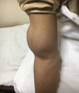
These tumours are more common than benign tumours of bone and can can occur at almost any site. They may or may not cause any symptoms such as pain and vary widely in appearance. Some of the commonest tumours are lipoma, fibroma, benign fibrous histiocytoma, schwannoma, hemangioma, giant cell tumor of tendon sheath and myxoma. Some of these may not need any further treatment and can be carefully observed after initial consultation and assessment by the sarcoma surgeon.
What are the worrying features?
If you have any of the following clinical features, you should seek a consultation to make sure you do not have a sinister pathology (cancer):
- Unexplained bone pain in any age.
- Pain at night.
- Pathological fracture (fracture without a significant injury).
- Lump bigger than 4 cm (bigger than a golf ball or table tennis ball).
- Lump increasing in size.
- Lump that is painful.
- Lump that is deep.
Investigations
Depending on the clinical features and type of lesion, your doctor may request a simple radiograph (X ray), Ultrasound, CT scan, MRI scan, Bone Scan or PET scan to get more information about the disease and its extent. With all this information in hand, you may need further investigation in the form of a biopsy or careful follow up.
Biopsy
This is a procedure where a sample of tissue is obtained from the suspected lesion (tumour) and sent to the histopathologist for it to be looked at under a microscope. Based on the results of this test, your doctor will discuss further treatment with you.
There are different types and techniques of biopsies and this depends on the nature and location of the tumour. Most soft tissue tumours will need a “truct biopsy”, where a sample is taken in clinic or a clean environment with a local anaesthetic with a very small incision. Occasionally, because of the location of tumour this sample may be taken with the help of ultrasound (Ultrasound guided biopsy).
For bone tumours, we usually perform core needle biopsies with a very small incision and x ray guidance in operating theatres as a day case procedure. If needed, they can be performed with a CT scan machine depending on the location of the tumour.
Sometimes the lesion is small, usually less than 2 cm, and your doctor will decide to perform an excisional biopsy. This involves taking the tumour out and then sending it for histology.
Surgery for bone tumours
Surgery for bone tumours depend on the nature and location of the lesion. In some instances (mostly benign bone tumours), the tumour can be scrapped out of the bone with or without the need for any further adjuvant treatment including bone graft or bone cementation. Depending on the size and location of defect, autograft (patient’s own bone) or allograft (bone from a bone bank) can be used to reconstruct the defect after removal of tumour.
In cases of larger defects after tumour excision, megaprosthesis (special joint replacements) are used to reconstruct the limb in order to preserve function and perform limb salvage surgery. These are complex reconstructions and your doctor will explain the procedure to you. Almost any part of bone or the whole bone with the involved joint can be replaced with these prosthesis. The advantage of this procedure is that patients can start mobilising almost immediately after surgery in comparison to bone grafts where the patient may have to wait for months before resuming function. The pros and cons of both of these reconstruction types will be explained to you by your doctor.
Depending on the nature of tumour and involvement of other important structures (including blood vessels and nerves), limb salvage surgery may not be possible every time and amputation of that limb may be needed to attain complete clearance from tumour. Advice regarding surgery and the type and procedure for arranging the prosthesis will be explained to you by your doctor. Counselling session will/can be arranged for you with our expert psychologist to alleviate all your concerns and guide you through this life changing event.
Surgery for soft tissue tumours
There are more than 50 different subtypes of soft tissue sarcomas which can arise in muscle, ligaments, fatty tissues, blood vessels and nerves. Following the confirmation of the nature nd type of sarcoma, your doctor will explain the treatment protocol to you.
Surgery, however, is the mainstay of treatment for soft tissue sarcomas. This is in the form of local excision of the tumour and may or may not involve a muscle flap or skin graft to cover the defect. Occasionally, a vascular bypass surgery is required if the blood vessel only is involved in the tumour. If however, both the important blood vessels and nerves are involved, limb salvage surgery may not be possible and ablative surgery will be required.
Depending on the nature and type of tumour pre or post-operative chemotherapy or radiotherapy may be needed. This will also depend on the results of histology after excision of the primary tumour.
Chemotherapy
Chemotherapy has transformed the whole treatment of sarcomas, particularly in malignant bone tumours. It has tremendously improved the survivorship of this select group of patients. It is used in isolation and sometimes in combination with radiotherapy to treat sarcomas, e.g: Ewing’s sarcoma. Following your biopsy results, the doctor may refer you to a medical oncologist for chemotherapy. In certain tumours, like osteogenic sarcoma, the patient is given 2-3 cycles of chemotherapy before surgery and 3-4 cycles after surgery depending on the aggressiveness of the tumour. All these decisions are made after discussion in a multidisciplinary team meeting.
The definite role of chemotherapy in the management of soft tissue sarcomas however still remains unclear and decisions are made about its utility depending on the type and nature of the tumour itself.
Radiotherapy
The role of radiotherapy also remains paramount in the management of sarcomas and depends on the nature and type of tumour. If needed, this is delivered by a radiation oncologist sometimes before surgery (e.g: in myxoid liposarcoma, Ewing’s sarcoma) or after surgery (depending on clarity of margins and aggressiveness of tumour).
Decision about the role of radiotherapy is made on an individual basis after discussion in a multidisciplinary team meeting
Follow up
Because of the nature of this pathology, patients are kept under follow up for a long time usually up to 10 years. Depending on the type of the tumour, the follow up maybe varied but it is usually every 3 months in the first 2 years, every 6 months thereafter till the 5th year and then annually. Clinical examination during the follow up visit is usually associated with check X- rays, CT scans or MRI scans as required. You may be asked to fill some questionnaires regarding your progress during the visits and this information is used for research and teaching purposes. However, all patient personal information is kept confidential and participation is not mandatory but very helpful for advancement of medical science and development of better treatment strategies for future.